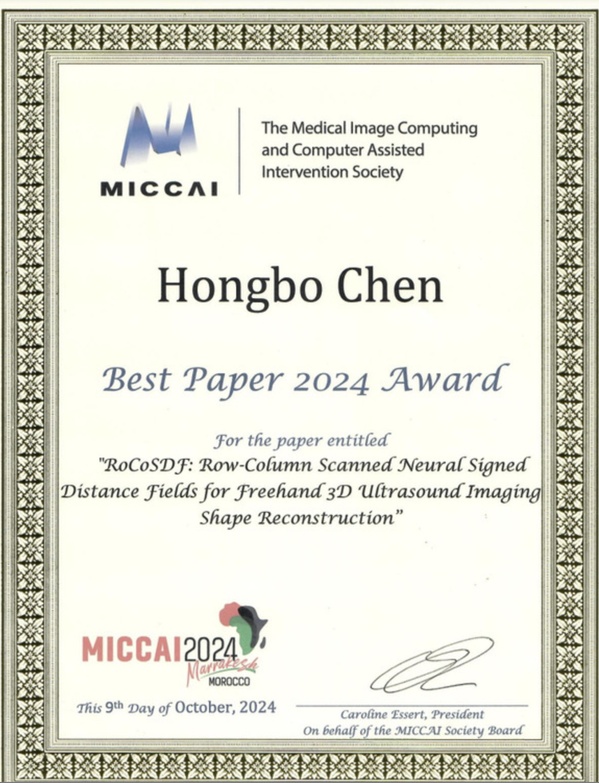
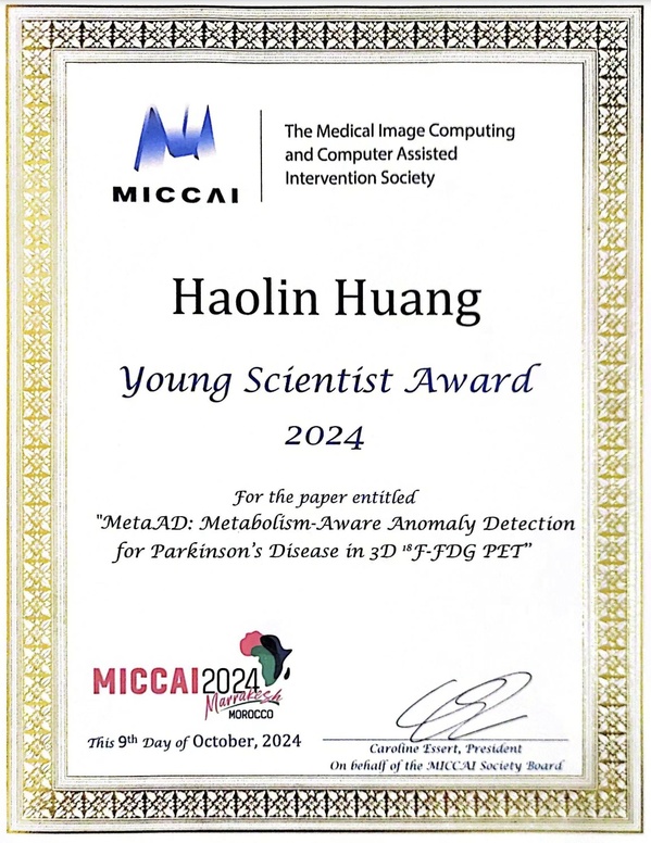
At the 27th International Conference on Medical Image Computing and Computer Assisted Intervention (MICCAI 2024), held from October 6 to 10, two students from ShanghaiTech University were honored for their outstanding research achievements in medical image computing and computer-assisted interventions. Chen Hongbo, a PhD student from Assistant Professor Zheng Rui’s group at the School of Information Science and Technology (SIST), received the “Best Paper 2024 Award” for his research on three-dimensional ultrasound multi-view shape reconstruction. Huang Haolin, a second-year master’s student from Associate Professor Wang Qian’s lab at the School of Biomedical Engineering (BME), won the “MICCAI 2024 Young Scientist Award” for his work on the accurately and efficiently detecting abnormal brain metabolic activity in patients with Parkinson’s disease.
In 2024, MICCAI received 2,879 paper submissions and accepted 857 papers from 43 countries and regions. The selection committee evaluated the papers and awarded one “Best Paper 2024 Award”, two Honorable Mentions, and five “Young Scientist Awards”, recognizing outstanding young researchers and highlighting the significance of their contributions in the field. Chen’s and Huang’s achievements reflect their strong research capabilities and underscore ShanghaiTech’s commitment to fostering innovative research and cultivating students.
The “Best Paper 2024 Award” was awarded for the paper entitled “RoCoSDF: Row-Column Scanned Neural Signed Distance Fields for Freehand 3D Ultrasound Imaging Shape Reconstruction.”
Traditional spinal surgeries, such as lumbar disc herniation procedures, often rely on intraoperative digital X-ray (DX) imaging to ensure accurate needle placement. Ultrasound-guided spinal surgery navigation can assist doctors locate targets in real-time while reducing radiation exposure for both doctors and patients. However, two-dimensional ultrasound is limited in displaying complete anatomical structures. Existing three-dimensional ultrasound imaging methods provide more spatial information but are constrained by ultrasound’s dependency on viewing angles and probe layer thickness, limiting their precision for surgical navigation.
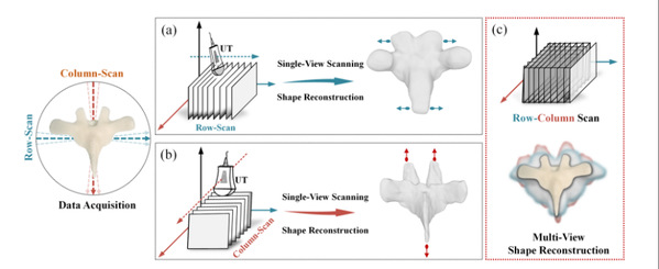
Two typical single-view scanning and reconstruction methods, and the multi-view scanning and reconstruction proposed in this study.
This study proposes a Row-Column scanned Neural Signed Distance Field (SDF), developing the first framework capable of accurately reconstructing three-dimensional anatomical structures from multi-view ultrasound scans. The framework uses four key technical steps to iteratively optimize the target structure from coarse to fine. This design overcomes the limitations of traditional pixel/voxel-level algorithms by enabling multi-view reconstruction to be directly edited and processed at the three-dimensional shape level.
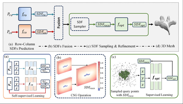
The three-dimensional ultrasound multi-view shape reconstruction framework proposed in this study.
The vertebral shapes reconstructed by this framework exhibit high geometric accuracy. The SDF constructed during the reconstruction process aids in real-time collision detection, providing early warnings if the surgical probe approaches the organ surface, thereby reducing surgical risks. Compared to the existing single-view reconstruction methods, the new framework significantly reduces the vertebral structure distortions caused by ultrasound view dependency and probe layer thickness. This improvement allows for more accurate and efficient target localization during surgery while reducing radiation exposure for both doctors and patients.
All six authors of the paper are from SIST: Chen Hongbo, a fifth-year PhD student; Gao Yuchong ’21, a second-year PhD student; Zhang Shuhang ’24, a first-year master’s student; Wu Jiangjie, a third-year PhD student; and their instructors, Assistant Professor Ma Yuexin from the Visual & Data Intelligence Center and Assistant Professor Zheng Rui from the Smart Medical Information Research Center. Prof. Zheng Rui is the co-corresponding author.
The “Young Scientist Award 2024” was awarded for the paper entitled “MetaAD: Metabolism-Aware Anomaly Detection for Parkinson’s Disease in 3D 18F-FDG PET.”
Parkinson’s disease (PD) is a common neurodegenerative disorder that primarily affects human’s motor system. Most hospitals lack access to specialized dopamine transporter (DAT) imaging technology for PD diagnosis, and the commonly used 18F-FDG PET imaging struggles to show major abnormalities of PD through visual analysis. The proposed Metabolism-Aware Anomaly Detection (MetaAD) framework addresses this challenge by converting input FDG PET images into synthetic CFT images with healthy patterns, and then reconstructing the FDG images using reverse modality mapping. This approach highlights metabolic abnormalities related to PD, with the visual differences between the input and reconstructed images serving as indicators of PD metabolic anomalies.
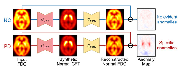
The MetaAD framework can highlight metabolic abnormalities of PDs in FDG images, with red and blue regions representing abnormal areas of higher and lower metabolism, respectively.
The MetaAD framework performed excellently in detecting metabolic anomalies of PD in 18F-FDG PET images, significantly improving the performance of computer-aided diagnosis (CAD) systems and helping radiologists diagnose PD more easily, efficiently, and accurately using 18F-FDG PET. Additionally, MetaAD holds potential for identifying new biomarkers, offering promising avenues for advancing PD research and treatment approaches.
This research was jointly completed by Huang Haolin; Shen Zhenrong, a PhD student from Shanghai Jiao Tong University; and Wang Jing, a PhD student from Huashan Hospital, Fudan University. Prof. Wang Qian and Prof. Zuo Chuantao from Huashan Hospital are the co-corresponding authors.

