An organoid is a miniaturized and simplified model of an organ created in vitro, designed to replicate the functional, structural, and biological complexity of that organ. In recent years, organoids have been widely used to study human tissues and organs. In general, the maturation of tissues and organs, as well as the normal functioning of the human body, depends on the crosstalk among different tissues. However, most current organoid models typically generate only a single tissue type. For instance, the interaction among tissues in the neuromusculoskeletal axis is particularly crucial. The neural, muscular, and skeletal tissues are closely related to each other in development, body function, and diseases. Modeling the human neuromusculoskeletal axis using organoids has proven to be a significant challenge.
Recently, the research group led by Assistant Professor Xiang Yangfei from the School of Life Science and Technology (SLST) at ShanghaiTech University constructed the first self-organized human neuromusculoskeletal organoids (hNMSOs) using human pluripotent stem cells. They provided an in vitro model for studying the human neuromusculoskeletal axis and related diseases. On December 9, their findings was published in a paper entitled “Generation of self-organized neuromusculoskeletal tri-tissue organoids from human pluripotent stem cells” in the journal Cell Stem Cell.

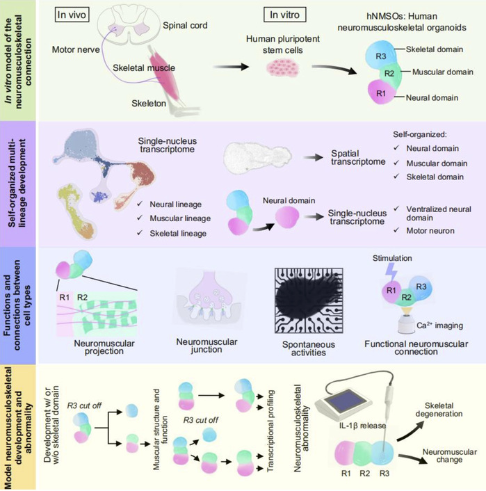
Unlike the previous attempts to generate complex organoids by assembling/fusing different organoids, this study employed a co-development strategy to generate hNMSOs from human pluripotent stem cells. This approach enabled the co-differentiation and spatial self-organization of the neural, muscular, and skeletal tissues, forming a composite organoid containing three tissues that self-organized into three regions while remaining interconnected.
Through immunostaining, single-nucleus transcriptome, and spatial transcriptome analyses, this study revealed the co-emergence and spatial self-organization of neural, muscular, and skeletal lineages in hNMSOs, effectively recapitulating human tissues. The neural domain of hNMSOs possessed the identity of the ventral spinal cord, and the researchers identified the generation of motor neurons therein through immunofluorescence staining and transcriptomics analyses. Methods such as retrograde tracing, multi-electrode arrays, optogenetic stimulation, and calcium imaging demonstrated the maturation and the functional connections among the tissues in hNMSOs. The study also showed that in hNMSOs, motor neurons in the neural domain can control the contraction of skeletal muscles in the muscular domain.
The researchers further utilized the hNMSOs model to analyze the importance of human skeletal tissue to the development of skeletal muscle. Through structural analysis, function research, and transcriptomics studies, the researchers demonstrated that the skeletal support in hNMSOs is beneficial to the development and maturation of human skeletal muscles. Additionally, this model was applied to study neuromusculoskeletal axis abnormalities in arthritis-associated conditions, revealing structural and functional abnormalities in neuromuscular connections following pathological skeletal degeneration.

This study marks the first successful construction of self-organized human neuromusculoskeletal tri-tissue organoids, providing an important model for exploring the interactions within the human neuromusculoskeletal axis, understanding the mechanisms of related diseases, and advancing drug discovery.
SLST PhD students Yin Yao, Zhou Wei, and Zhu Jinkui from Prof. Xiang’s group are co-first authors of this paper. Prof. Xiang is the corresponding author, and ShanghaiTech University is the primary affiliation.
*This article is provided by Prof. Xiang Yangfei

