The International Society for Magnetic Resonance in Medicine (ISMRM) 2025 Annual Meeting will be held from May 10 to 15. During the call for papers for ISMRM 2025, the School of Biomedical Engineering (BME) at ShanghaiTech University achieved remarkable success, with 43 submissions accepted, comprising 10 oral presentations and 2 power pitch presentations. The exceptional outcome underscores BME’s research strength and innovative capacity in the field of magnetic resonance, while also reflecting its sustained progress and promising potential for further advancements in related domains.
Established in 1994, ISMRM is the largest and most authoritative international professional society in the field of magnetic resoance, with over 8,500 members, including clinicians, physicists, engineers, biochemists, and technical experts from various disciplines. The society aims to promote the research, development, and application of magnetic resonance technology in the fields of medicine and biology. The ISMRM annual meeting is a top-tier academic event in this field. In 2025, the conference received 10,452 abstracts, with only 7.6% selected for oral presentations, 524 for power pitch presentations, 3,241 for digital posters, and 554 for traditional posters.
BME’s representative achievements accepted by ISMRM 2025
Below are highlights of research works from various research groups accepted for ISMRM 2025:
General Multi-modal MRI Synthesis with Metadata-guided Text Encoder
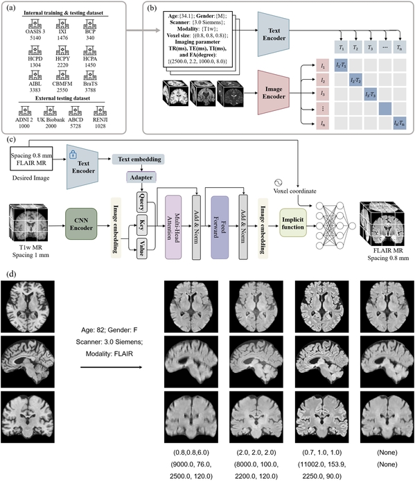
In this research, Wang Yulin, a postdoctoral researcher in Professor Shen Dinggang’s group, proposed a pioneering general multi-modal MRI synthesis framework guided by metadata for multi-modal magnetic resonance imaging synthesis. This framework is capable of generating high-quality MRI sequences based on specified scanning parameters and demonstrates excellent cross-center generalization capabilities. It offers a new solution for quickly and cost-effectively obtaining customized multi-modal MRI images, expected to significantly enhance the efficiency and precision of clinical diagnosis and research.
BrainParc: Unified Lifespan Brain Parcellation with Anatomy-guided Progressive Transmission
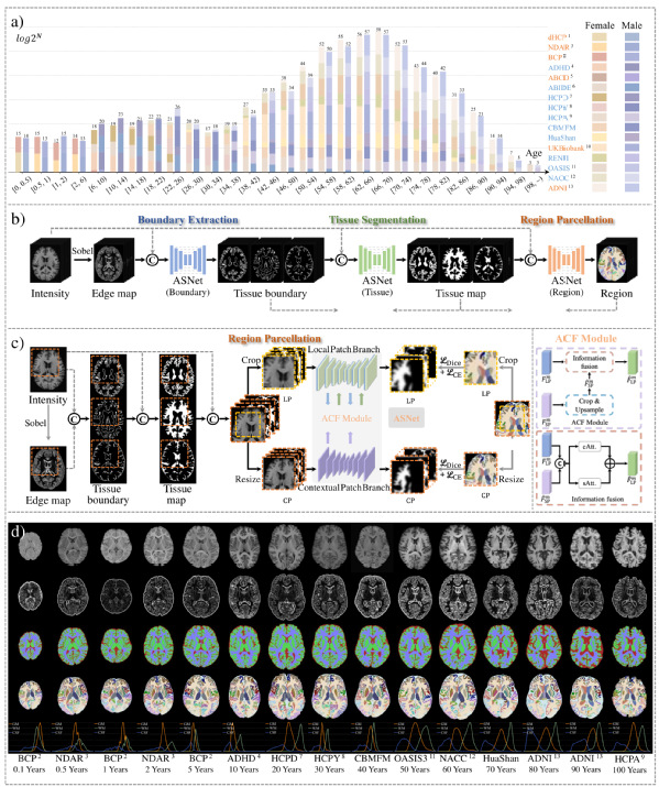
PhD student Liu Jiameng in Prof. Shen Dinggang’s group proposed an automated and robust framework for brain tissue segmentation and parcellation across the entire human lifespan. The framework can process structural MRI data from any age, MRI instrument, and scanning parameters, providing new possibilities for large-scale exploration of human brain development.
Simultaneous Myocardial T1, T2, T1ρ and Fat Fraction Mapping with Hybrid Dual-echo Cartesian Acquisition and Dictionary Matching
PhD student Lü Zhenfeng in Assistant Professor Qi Haikun’s research group developed a new MRI technique called “Dixon-MultiMap,” which enables the simultaneous and precise quantification of cardiac fat fraction and T1/T2/T1ρ parameters. This technique integrates hybrid multi-echo Dixon readouts with a multi-parameter quantitative framework, using four-echo data to obtain high-precision B0 field and fat fraction maps while improving the accuracy of water-fat separation in dual-echo multi-contrast images. The water-phase multi-contrast images are then matched with a dictionary to achieve B1+-corrected multi-parameter quantification. This technique provides an efficient and integrated solution for the non-invasive assessment of fat-infiltrative myocardial diseases.

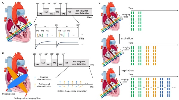
A Fast 2D Free-breathing Myocardial T1 Mapping Technique with Multi-slice Free-running Acquisition and Self-navigated Motion Calibration
Master’s student Huang Hongzhang in Prof. Qi Haikun’s research group proposed a retrospective through-plane motion correction scheme to address respiratory motion problem during MRI scanning, enabling T1 quantification during free breathing. The study introduced a multi-slice joint scanning and self-navigated self-calibration module, allowing for respiratory signal extraction and through-plane motion correction without external detection devices, offering new insights into cardiac MRI motion correction.
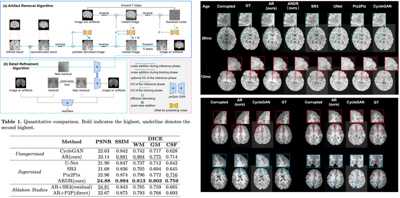
Unconditional Diffusion Model for 3D MRI Artifact Removal and Detail Refinement
In this study, Deng Haowen and Zhu Zihao, master’s students in Associate Professor Zhang Han’s research group, proposed an unconditional diffusion-based algorithm for removing head motion artifacts in MRI scans of children aged from 0 to 6 years. The framework only uses artifact-free data for training and inference, overcoming the reliance on paired datasets required by traditional artifact removal algorithms.
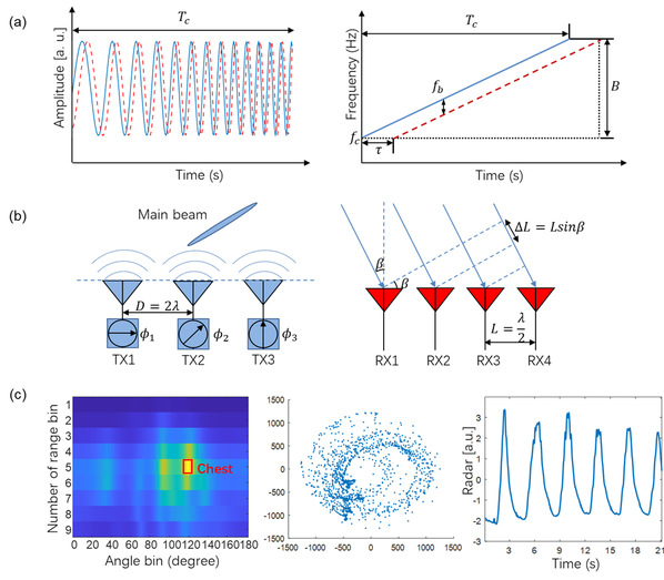
Frequency Modulated Continuous Wave Radar-based Non-contact Prospective Respiratory Motion Estimation and Correction
Research Assistant Diao Jiameng in Professor Hu Peng’s research group proposed a millimeter-wave radar-based prospective respiratory motion correction technique for cardiac MRI. The technique enables real-time estimation and prospective correction of respiratory motion during MRI data acquisition, overcoming the limitations of traditional respiratory gating. This technology provides a new solution for free-breathing cardiac MRI scans, compatible with various clinical sequences such as cardiac cine imaging and myocardial quantification.
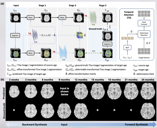
A Deep Learning Approach to Longitudinal Infant MRI Synthesis
In this research, master’s student Fang Yu in Prof. Wang Qian’s research group proposed a multi-stage longitudinal synthesis framework for brain MRI images of infants aged 0-24 months. The innovative framework can synthesize high-fidelity infant brain MRI images at any given age, providing new technical means for constructing a comprehensive longitudinal dataset of infant brain development and potentially supporting research on early human brain development.
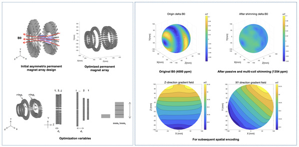
Design of Asymmetric Permanent Magnet Arrays for the Development of a Portable MR Head Imager
Shao Zejie, a master’s student in Assistant Professor Ren Zhihua’s research group, proposed an innovative asymmetric permanent magnet array design, opening new technical pathways for the development of low-field portable MRI systems. By optimizing algorithms to overcome size limitations of traditional arrays, this asymmetric design significantly enhances magnetic field performance. The team also pioneered the inclusion of magnetic field harmonic components in portable MRI evaluation systems, achieving precise control of field uniformity and high-order harmonics using passive shimming technology.
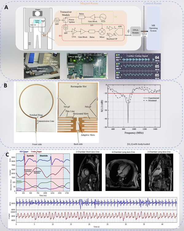
Cardiac MR Gating Using a Non-contact MR-compatible Doppler Radar System at 3T: Initial Results on Healthy Subjects
Backed by the United Imaging Research Institute and United Imaging Healthcare (UIH), this collaborative study from the research groups of Professors Ren Zhihua and Hu Peng seamlessly integrates intelligent analysis modules and cutting-edge cardiac gating algorithms into a next-generation MRI-compatible cardiac radar system, co-developed by BME and UIH. This innovative system enables high-precision, low-latency detection of millimeter-scale cardiac signals for MRI gating. Unlike traditional ECG-based gating, this non-contact approach enhances patient comfort, mitigates risks associated with conventional contact methods, and offers a safer, more dependable solution for the future of cardiac magnetic resonance imaging.
4D Cardiac Shape Reconstruction Using Image-Aided Neural Signed Distance Correction Fields
In this study, Zhang Zichen, a visiting student in both Assistant Professor Cui Zhiming’s and Assistant Professor Zhang Zeng’s research group, used explicit semantic encoding based on cardiac MRI images to guide neural networks in learning continuous myocardial motion. The method uses continuous signed distance fields to describe myocardial shape and achieves high-quality myocardial reconstruction. Explicit semantic encoding improves reconstruction accuracy and accelerates network convergence. This study provides a foundation for subsequent cardiac blood flow simulations.

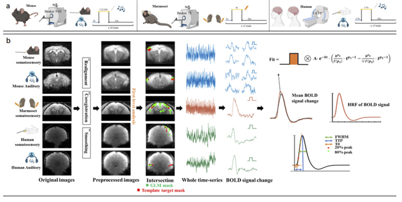
Characterizing Hemodynamic Responses Across Species in the Awake State
In this study, master’s student Chen Qian in Assistant Professor Ma Zhiwei’s research group systematically compared the spatiotemporal characteristics of hemodynamic response functions in mice, marmosets, and humans in the awake state. This research provides key parameters for the translational application of neuroimaging animal models.