Recently, researchers from the Center for Transformative Science (CTS) at ShanghaiTech University achieved an exciting advancement: they used an ultrafast X-ray pulse to capture the internal structure of a two-dimensional (2D) crystal made of gold nanoparticles in a single shot, like taking an instant photo. Published in IUCrJ with title “Single-shot X-ray imaging of two-dimensional strain fields in colloidal crystals,” this study not only reveals defects inside the crystal but also paves the way for exploring how materials change in mere fractions of a second.
Colloidal crystals: tiny particles building amazing structures
Ever wondered how particles just tens of billionths of a meter wide can stack up neatly like building blocks? That’s what colloidal crystals are—materials formed when tiny particles arrange themselves into orderly patterns. This time, the team studied a 2D colloidal crystal, a flat “map” of particles. Scientists are keen on these crystals because they help us understand how materials bend, break, or even melt, and they might one day improve things like batteries or light-based devices. To create this crystal, the team used gold nanoparticles about 40 nanometers in size, dropped them onto Si3N4 membranes, and let the water evaporate, leaving the particles to form a crystal on their own.
X-ray single-shot imaging: seeing inside with one flash
So, how do you peek inside such a tiny crystal? The researchers turned to a technique called Bragg coherent diffraction imaging (BCDI), which uses X-rays to “see” a crystal’s structure, especially its imperfections. They fired an ultrafast X-ray pulse from the Shanghai Soft X-ray Free Electron Laser Facility (SXFEL). This pulse, lasting just 100 femtoseconds (a femtosecond is a trillionth of a second), hit a 3-micron spot and captured the crystal’s full structure in one go. Though this powerful flash destroys the crystal, it’s so quick that it grabs the details before the damage happens.
In the experiment, the X-ray beam struck the gold nanoparticle crystal, producing a hexagonal diffraction pattern. These patterns revealed the crystal’s hidden story: some areas had misaligned particles, showing cracks and gaps. To double-check, the team had already looked at the crystal with an electron microscope, and the X-ray results matched up perfectly.
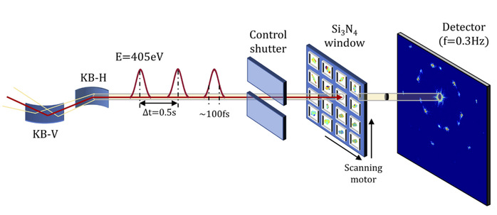
Diffraction beamline layout of the experiment at the Coherent Scattering and Imaging (CSI) beamline of the SXFEL.
From patterns to maps: decoding the defects
By analyzing the X-ray diffraction patterns, the team created 2D “displacement maps” and “strain tensor maps.” In simple terms, displacement maps show how much the particles shifted from their ideal periodic positions, while strain tensor maps highlight where the crystal was stretched or squeezed quantitatively. They found clear zones inside the crystal where defects stood out like boundaries. This imaging was so precise it reached resolutions as fine as 110 nanometers.
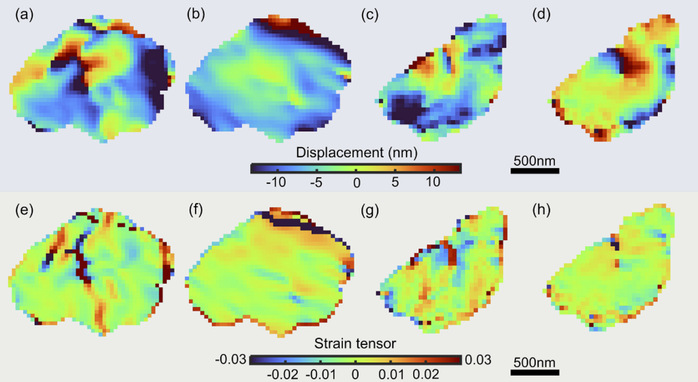
Atomic displacement maps (a-d) and strain tensor maps (e-h) of two crystals reconstructed from single-pulse diffraction patterns after phase retrieval.
Why it matters
This study doesn’t just give us a clearer picture of colloidal crystal structures; it also proves that X-ray single-shot imaging can unlock the secrets of 2D materials. “With this technique, we can see the tiniest flaws in a crystal instantly, opening doors to understanding how materials behave under extreme conditions,” said Professor Jiang Huaidong, one of the lead researchers. Looking ahead, the researchers want to add a laser “pump” to see how the crystal melts ultrafast when struck by a laser. This could lead to better materials for technology and deeper insights into the rules of the 2D world.
Research Assistant Professor Diao Jiecheng and Postdoctoral Researcher Gao Zichen from the CTS are the co-first authors of the paper. Professor Jiang Huaidong and Research Professor Fan Jiadong from the CTS, and Professor Ian Robinson from University College London are the co-corresponding authors.